
Introduction
Pregnancy is a beautiful and miraculous journey for any woman, but it often comes with various concerns and considerations, especially for women over the age of 35. Advanced maternal age, commonly defined as being 35 years or older at the time of delivery, is associated with increased risks for both the mother and the baby. One essential aspect of prenatal care for women in this age group is the use of ultrasound examinations to monitor the development and well-being of the fetus.
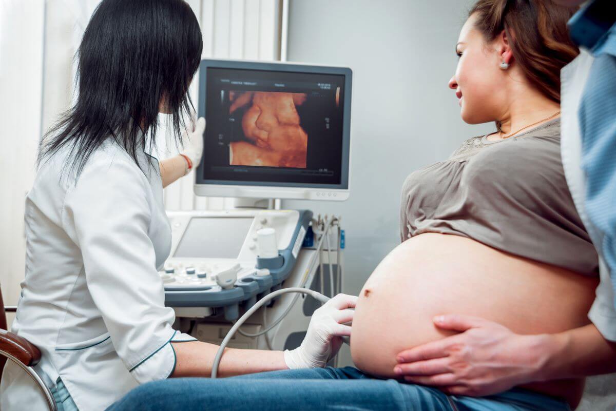
Ultrasound imaging is a valuable tool in obstetrics, allowing healthcare providers to visualize the fetus, assess its growth, monitor the placenta, and detect any potential abnormalities or complications. However, the frequency of ultrasound examinations during pregnancy varies depending on several factors, including maternal age, medical history, and any existing risk factors.
For women over 35, healthcare providers may recommend more frequent ultrasound scans compared to younger mothers. This is because advanced maternal age is associated with a higher incidence of certain pregnancy complications, such as gestational diabetes, preeclampsia, and chromosomal abnormalities like Down syndrome. Additional ultrasound screenings can help detect these issues early on, allowing for timely interventions and appropriate management.

Is 35 years old a high risk pregnancy?
After age 35, there’s a higher risk of pregnancy-related complications that might lead to a C-section delivery. The risk of chromosomal conditions is higher. Babies born to older mothers have a higher risk of certain chromosomal conditions, such as Down syndrome.
At the age of 35, pregnancy is generally considered to be of advanced maternal age (AMA). While it doesn’t automatically classify as high-risk, it does carry some increased risks compared to pregnancies in younger women. One of the main concerns is the higher likelihood of chromosomal abnormalities, such as Down syndrome, due to the aging egg quality. Additionally, women over 35 are at a higher risk for gestational diabetes, high blood pressure, and pregnancy complications such as placenta previa and preeclampsia.
Despite these risks, many women over 35 have healthy pregnancies and deliveries. It’s important for older expectant mothers to receive thorough prenatal care, including regular check-ups, screenings, and tests to monitor for any potential complications. With proper medical attention and management, many women over 35 go on to have successful pregnancies and healthy babies.
How many ultrasounds during pregnancy over 35?
Women over 35 may need to be monitored more closely during pregnancy depending on any pre-existing health conditions they have. This would mean more ultrasounds (2-3), more tests to check for heart diseases or gestational diabetes, and non-stress tests in the final weeks of the pregnancy.
The number of ultrasounds during pregnancy for women over 35 varies depending on individual circumstances and healthcare provider recommendations. However, it’s common for women in this age group to undergo the same standard prenatal ultrasound screenings as younger women, typically including an early dating scan in the first trimester, an anatomy scan around 20 weeks, and possibly additional ultrasounds if there are specific concerns or complications.
Women over 35 may be offered specialized screenings such as non-invasive prenatal testing (NIPT) or amniocentesis to assess for chromosomal abnormalities, given the increased risk in this age group. These screenings are typically offered in conjunction with, rather than in place of, standard ultrasounds.
How many ultrasounds during pregnancy over 40?
Women over 40 with risk factors may receive ultrasounds every 4 to 6 weeks. Potential prenatal DNA screening, genetic counseling or diagnostic testing. Antenatal fetal surveillance: tracing heart rate, fetal activity and amniotic fluid (once or twice per week).
Similarly to women over 35, the number of ultrasounds during pregnancy for women over 40 depends on various factors such as medical history, risk factors, and healthcare provider recommendations. However, due to the increased risks associated with advanced maternal age, women over 40 may undergo more frequent monitoring and ultrasounds compared to younger expectant mothers.
In addition to the standard prenatal ultrasound screenings typically offered to all pregnant women, such as the dating scan and anatomy scan, women over 40 may be offered additional screenings and tests to assess for chromosomal abnormalities and other potential complications. This may include non-invasive prenatal testing (NIPT), amniocentesis, or more frequent ultrasounds to monitor fetal growth and well-being.
How many times can you do ultrasound during pregnancy?
Most healthy women receive two ultrasound scans during pregnancy. “The first is, ideally, in the first trimester to confirm the due date, and the second is at 18-22 weeks to confirm normal anatomy and the sex of the baby,” explains Mendiola.High-risk pregnancies often require more frequent monitoring, including a higher number of ultrasounds compared to low-risk pregnancies.
The specific number of ultrasounds for a high-risk pregnancy varies based on individual circumstances, medical history, and the presence of specific risk factors. Conditions such as gestational diabetes, hypertension, or multiple gestations may necessitate more frequent ultrasounds to monitor fetal growth, assess placental function, and evaluate amniotic fluid levels.
Pregnancies complicated by factors like maternal age, pre-existing medical conditions, or a history of pregnancy complications may require closer surveillance with increased ultrasound frequency. The goal of more frequent ultrasounds in high-risk pregnancies is to detect and manage potential complications promptly, ensuring the health and well-being of both the mother and the fetus.

Is 4 ultrasounds too many?
“Pregnant women can take a sigh of relief knowing that there is no evidence showing that ultrasound is harmful to a growing baby at any stage of pregnancy.” Whether four ultrasounds are too many depends on several factors, including the specific circumstances of the pregnancy and the medical necessity for each scan. In many cases, four ultrasounds may be considered appropriate and necessary for ensuring the health and well-being of both the mother and the fetus.
The first trimester typically involves two ultrasounds: a dating scan to confirm the pregnancy and determine gestational age, and a nuchal translucency scan to assess the risk of chromosomal abnormalities. The anatomy scan in the second trimester provides a comprehensive evaluation of the baby’s development and is an essential part of routine prenatal care. If additional ultrasounds are recommended later in pregnancy due to specific concerns or risk factors, such as monitoring fetal growth or assessing amniotic fluid levels, these scans may be justified in providing essential information for clinical management.
The decision to perform multiple ultrasounds should be made based on individual medical needs and the judgment of the healthcare provider. While there is no universally agreed-upon limit to the number of ultrasounds, it’s essential to ensure that each scan is medically justified and necessary to avoid unnecessary exposure to ultrasound waves.
How many ultrasounds for high-risk?
You will have at least two ultrasounds during your early and middle pregnancy, and in the later parts of your high-risk pregnancy, you may have ultrasounds as often as once a week based on your health needs and situation. High-risk pregnancies often require more frequent monitoring, including a higher number of ultrasounds compared to low-risk pregnancies.
The specific number of ultrasounds for a high-risk pregnancy varies based on individual circumstances, medical history, and the presence of specific risk factors. Conditions such as gestational diabetes, hypertension, or multiple gestations may necessitate more frequent ultrasounds to monitor fetal growth, assess placental function, and evaluate amniotic fluid levels.
pregnancies complicated by factors like maternal age, pre-existing medical conditions, or a history of pregnancy complications may require closer surveillance with increased ultrasound frequency. The goal of more frequent ultrasounds in high-risk pregnancies is to detect and manage potential complications promptly, ensuring the health and well-being of both the mother and the fetus.
How many ultrasounds are done in the 3rd trimester?
More and more obstetricians refer women for two third trimester ultrasounds, mostly at 28 weeks and 36 weeks, because there is increasing evidence that this allows better detection of growth problems and ensures better outcomes for babies.In most pregnancies, routine ultrasound screenings are not typically performed during the third trimester unless there are specific medical indications or concerns that warrant additional monitoring.
While the number of ultrasounds in the third trimester can vary based on individual circumstances, many pregnancies may not include any ultrasound examinations during this time. However, some healthcare providers may recommend additional scans in the third trimester for various reasons, such as assessing fetal growth, measuring amniotic fluid levels, or monitoring for signs of complications like placental abnormalities or fetal distress.
The decision to perform ultrasounds in the third trimester is individualized and based on the specific needs and concerns identified during routine prenatal care. Healthcare providers will determine the appropriate frequency of ultrasounds in the third trimester to ensure the health and well-being of both the mother and the baby.
Recommended ultrasounds for over-35 pregnancies?
Given increased rates of multiple gestations for pregnant individuals with anticipated delivery at age 35 years or older, we suggest a first-trimester ultrasonogram.Pregnancies in individuals over the age of 35 are often considered advanced maternal age and may be associated with an increased risk of certain complications. While there is no set number of ultrasounds recommended specifically for over-35 pregnancies, routine prenatal care typically includes the same standard ultrasound screenings as for younger pregnancies.
These screenings may include a first-trimester dating scan to confirm the pregnancy and determine gestational age, as well as a second-trimester anatomy scan to assess the baby’s development. Additionally, healthcare providers may offer additional screenings such as non-invasive prenatal testing (NIPT) or amniocentesis to assess for chromosomal abnormalities due to the increased risk associated with advanced maternal age.
The frequency and timing of ultrasounds for over-35 pregnancies should be determined on an individual basis in consultation with the healthcare provider, taking into account factors such as medical history, overall health, and any specific concerns identified during routine prenatal care.

Conclusion:
Prenatal care for women over 35 often involves more frequent ultrasound examinations to monitor the health and development of the fetus. These additional screenings play a crucial role in detecting potential complications early on, allowing for timely interventions and improved outcomes for both the mother and the baby. While advanced maternal age may pose certain risks during pregnancy, modern medical technology and advancements in obstetric care help ensure the best possible outcomes for women in this age group. By working closely with their healthcare providers and adhering to recommended prenatal screening protocols, women over 35 can navigate their pregnancy journey with confidence and peace of mind, knowing that they are receiving the care and support they need to welcome a healthy baby into the world.